Archive

Pall Mall Medical PACS Migration

Pukka-j recently relocated the RIS / PACS services at Pall Mall Medical to new hardware at a different site.  Pall Mall Medical were keen to bring the archiving services in-house and redirect all the modalities to the new service.  Due to limited access and network bandwidth where the data was originally located, Pukka-j worked closely with the IT partners at Pall Mall (Ascent Networks) to find a way to move the services whilst minimising the impact to the department.  The process was made simpler because it involved transferring data and services between two Pukka-j systems.

The initial stage was to set up the new services on the new hardware and start to establish connectivity with modalities and external DICOM and HL7 services like teleradiology companies.  In order to move the data, the simplest way was to export it to an encrypted external drive and simply copy the data from the old system to the new one along with all the records in the database.  Importing the database records brought the new system up to date but there was a small delay in the department whilst all the DICOM data was restored.

Peter Charlton, Senior IT Support at Ascent Medical told us “The product and support we receive from Pukka J are second to none. The internal knowledge of the systems allows Pukka J to provide us with quick and efficient support to any issues with have.”

Find Out More

Please contact us to find out more about our PACS and RIS solution or request an online demonstration

Find Out More


Radiology Teaching Archive at Queen Margaret University

Pukka-j recently installed a new radiology teaching archive at Queen Margaret University to help the department deliver teaching images to radiography students.

A windows server has been configured with the Pukka-j teaching archive to provide access to example radiology images to students.  The images are being sent to the archive via a Pukka-j Nexus service running in the hospital network with a DICOM connection to the hospital PACS.  The Nexus service has been configured with the anonymisation plug-in such that any identifiable information is removed from the DICOM header of the images as they pass through.  Because of the streaming nature of the Nexus service, the images are not cached locally and pass though the service, being modified as they are transferred.  In addition to the anonymisation plug-in, the Nexus service has been setup with the redaction plugin so as to remove any demographics in the banner from any ultrasound images and also from the report screen captures of the Dexa images.  DICOM SRs also pass through the service and are attached to the study on the teaching archive.  These SRs are reviewed at the university, to remove any information that is not required before being made available as part of the study.  The images show how the Dexa screen captures have the patient information removed during redaction.

Dexa_Before

Dexa_After

Find Out More

Contact us to find out more about our software or to arrange an online demonstration

Find Out More

Pukka-j delivers unique DICOM storage solution for Cookridge Hospital, Leeds teaching hospitals NHS Trust

At Cookridge Hospital, Pukka-J delivered a DICOM store designed specifically for Oncology workflow as part of the Leeds Clinical Treatment System (CTS).

When looking for a system to handle, store and transfer oncology data, Cookridge Hospital wanted to take the opportunity to develop and improve existing radiotherapy systems and be able to store all images, plus RT objects including RT plans and RT structures, in a single central location. They required robust permanent and semi-permanent storage, with the possibility of evolving into a future PACS system. In addition, it should be ‘future proof’, fully DICOM-compliant, and also compliant with the emerging NHS information requirements (NPfIT/Connecting for Health).
One of the known difficulties with Radiotherapy Treatment Planning is system integration, since some of the devices are not fully DICOM compliant. Pukka-J met the challenge by providing the DICOM infrastructure for a central repository for radiotherapy images to be stored, and at the same time dealt with the different DICOM characteristics across the mix of vendors. The intelligent DICOM Store is capable of facilitating the centralised storage and also of sharing oncology data across a heterogeneous mix of radiology and radiotherapy modalities. The Store can handle DICOM radiotherapy objects, a mix of radiology DICOM images, while at the same time importing DICOM images from a local network, as well as importing DICOM images from referring hospitals.
Bob Wheller, Head of Radiotherapy Technology Services, Cookridge Hospital, Leeds Teaching Hospitals NHS Trust says:

We selected Pukka-J because we felt we would get value for money with cost effective DICOM storage. In addition the Company was extremely flexible and had the ability to develop the system within the stipulated timescales. With a thorough grasp of the complexity of radiotherapy services, Pukka-J responded quickly to problems we encountered.

In fact, there are three separate stores. Firstly, an Image Import Store for remote images to be sent or copied from a referring hospital CD. This Store contains a quarantine area which deals with any conflicts with patient or hospital demographics’ information, filtering and modifying such information prior to entering the second Store.
The second store is the “Radiotherapy Planning Store – a temporary repository for the Treatment Planning Department, and described by Cookridge as a big patient folder since it is used to automatically transfer radiotherapy and radiology objects backwards and forwards between mixed manufacturer radiotherapy planning modalities. Images are automatically routed between the processes and computers that perform the RT services such as treatment planning and treatment delivery.
The third and final store is the Main Store for the permanent storage of all images and valid RT objects that are used in the actual treatments. It holds the patient’s data after the course of treatment, and is held ready for PACS storage, where it can be easily integrated or migrated into any major PACS system.
As a safety net, Pukka-J also delivered a resilient back-up and redundancy to the DICOM Store. Using the Pukka-J’s DICOM mirror software, data is automatically backed-up and provides a unique system – should any one unit fail, the mirrored DICOM Server prevents loss of data and workflow as everything is totally online and easily accessible.

By |Archive|Comments Off on Pukka-j delivers unique DICOM storage solution for Cookridge Hospital, Leeds teaching hospitals NHS Trust

Pukka-j responds to demand for a solution to upload CD images to online facility

Pukka-j has developed a unique Medical Disk Service (MDS) specifically designed for Nuclear Medicine and PET/CT departments to enable import or export of DICOM compliant CD or DVD removable media. To develop this new device, Pukka-j partnered with Rimage Corporation, the world’s largest and most experienced manufacturer of CD and DVD publishing, duplication and printing systems, providing state-of-the-art equipment, representing the very latest advances in CD and DVD publishing technology.

The device itself features an input bin for stacking multiple CD and DVD’s into one compartment of the MDS robot. The robot then takes one disk after the other from the stack, reads the image data ready for the department to view, store and manage. The system is so quick and easy that the stacks of CDs collecting dust on shelves could be a thing of the past in Nuclear Medicine departments.
The MDS, customised by Pukka-j, enables a completely web-based solution that can be accessed via any networked PC. It intelligently manages all imported images via the Pukka-j DICOM Server by automatically sorting the imported images from the CD or DVD into multiple folders based on any DICOM field complete in the image header. This could be modality type, body part, institution or referring site. For instance, all PET CT image CDs acquired from mobile ISTC Service ‘A’ will be stored in one specific folder ready for image archive or distribution.

The system comes complete with an in-built short-term image cache server and can be easily expanded to feature Networked Attached Storage of up to several TB’s of RAID for long-term storage. It is also well suited for departments who wish to keep raw PET/CT and NM data separate from radiology images, where often only screen captures are relevant to the radiology PACS/RIS. In addition, the imported image’s unique identifier can be prefixed by Pukka-j to enable the user group to identify, quickly and easily, all imported images and keep the data separate, but accessible from PACS workstations.
Another feature is Pukka-j’s inbuilt NM and PET CT connectivity which allows imported images to be viewed and manipulated on existing specialist Philips, Siemens and GE workstations.
The MDS image distribution is also well catered for with Pukka-j’s Advanced DICOM Rules for auto-forwarding data across the network. A typical rule could be to send all PET CT images to workstation A, B and C except head images, which can only be sent to workstation D.
For research and education requirements, an automated rule to remove the data and depersonalise the image can be applied to any image data, to remove patient, referrer and institution identification. The de-personalised data can then be auto-routed to a specified location or DICOM device/node via Pukka-j’s advanced Server rules.

By |Archive|Comments Off on Pukka-j responds to demand for a solution to upload CD images to online facility

Pukka-j offers a ready made solution for CD Password Protection and Encryption

Pukka-j is able to offer a ready-made solution for encryption and password protection that provides users with a flexible range of products, depending on the throughput of discs required by an organisation. For many years now, the Pukka-j product portfolio has contained encryption, password protection and anonymisation techniques, as essential tools for the user, enabling the confident distribution of discs as and when needed. One of the methods of sharing image data is to export images from a PACS or direct from an imaging modality to a CD or DVD for image-sharing distribution. Due to the recent Government Data Handling Review report, there is a mandatory requirement that all removable media, including CD and DVD containing patient data, must be password protected and encrypted for distribution.

Kevin Wilson, Managing Director of Pukka-j says:

Prior to the roll-out of Virtual Private Networks between Hospital and on-call A&E reporters, Pukka-j very early on utilised data protection methods for safeguarding sensitive details for our systems. This allowed images to be sent outside of the hospital network and across the internet, as the transmission of data across the internet required strict adherence to data security policies. As a company, we have always valued encryption, password protection and anonymisation, as user-essential tools for providing the assurances of data security within our product range.

For organisations requiring a high output of burning images to discs, Pukka-j is able to offer a full range of high-performance Rimage-manufactured publishing devices. The Pukka-j Medical Disk System (MDS) is a fully-integrated range of CD/DVD robots that require no additional PC to drive the process; the system can be connected to an imaging network, allowing the MDS to be accessed from anywhere on the network, and to be easily added as an existing PACS accessory. The Pukka-j system Administrator User Interface allows automatic encryption and password protection for all data being ordered to disks as a default setting, requiring no extra click from the user.

By |Archive|Comments Off on Pukka-j offers a ready made solution for CD Password Protection and Encryption

Pukka-j provides novel solution for Brighton and Sussex University Hospitals NHS Trust

Now up and running, the Pukka-J solution for image viewing, archiving and distribution is fulfilling a key role within the Trust. At the same time the archiving solution provided to the Royal Sussex County Hospital prepares the Hospital for PACS, with all CT, MRI PET and NM image being stored direct to the Trust’s existing SAN. The Trust will be deploying NPfIT PACS in the very near future and the Pukka-J storage system will be utilised for specialist areas such as Oncology and On-call reporting.

At the Royal Sussex, Pukka-J’s web browser product for teleradiology: Dicom Explorer is used primarily for the distribution and display of medical images (CT, MRI PET and NM), for Radiology, Oncology, A&E, Neurological Services and Renal Medicine Departments. Images can also be shared with other hospitals within the Trust, including the Sussex Cancer Centre and Princess Royal Hospital, Haywards Heath. Pukka-J’s PACSPost allows on-call consultants to pick up images from a secure site immediately, view remotely and provide a 24/7 “on call” reporting service, including emergency reporting on CT and MRI images.

Dr. Graham Dodge, Lead Consultant Radiologist at the Royal Sussex Hospitals says:

We have been particularly pleased with the image distribution facility, how fast it is, and the fact that my radiological colleagues and I can report on a variety of images from anywhere in the hospital. It also allows clinicians to access images from their clinics and offices. Using PACSPost allows us to view on call-scans securely and reliably from our homes.

The Sussex Cancer Centre, situated in the Royal Sussex County Hospital , Brighton, using Pukka-J’s system, is also able to view images from the Agfa PACS at Eastbourne District General Hospital and The Conquest Hospital, Hastings . Next in line, is a link to images from the Ferrania PACS at Worthing and Southlands Hospital , Worthing . The Centre is a key specialist cancer care organisation providing oncology treatment within the Sussex cancer network area. It serves a population of just over one million people, covering an area stretching from Hastings in East Sussex to Worthing in West Sussex . Hospitals within the network include The Royal Sussex County Hospital, Brighton Royal Hospital , Hayward ‘s Heath Hospital , Worthing and Southlands Hospital , Eastbourne DGH and the Conquest Hospital in Hastings . The main function of the Centre is the provision of both radiotherapy and chemotherapy treatment; the fast and easy access to images via the Dicom Exporer is vital for the efficient and timely diagnosis and treatment of patients.

By |Archive|Comments Off on Pukka-j provides novel solution for Brighton and Sussex University Hospitals NHS Trust

Winner of Pukka-j’s prize at BNMS Spring Meeting 09

The winner of Pukka-j’s prize draw at the BNMS meeting in Manchester was Elaine Woods, Trainee Clinical Scientist at the Royal Free Hospital. The prize, which was an iPod player, was presented to Elaine by Dez Wright, Software & Applications Physicist at Pukka-j. The winning entry was drawn by Dr. Rak Ganatra, Chairman of the Scientific BNMS Committee and Consultant Radiologist, Nuclear Medicine Physician & PETCT Specialist at Nottingham University Hospitals.


Shown here are Louise Crossley
(Pukka-j Business Development Director)
with Dr. Rak Ganatra making the draw.

Shown here are Dr Dez Wright
(Pukka-j Software & Applications Physicist)
presenting the prize to Elaine Woods.

By |Archive|Comments Off on Winner of Pukka-j’s prize at BNMS Spring Meeting 09

Addenbrooke’s Hospital Cambridge selects Pukka-j’s Oncology Store for its Oncology Centre

Download this article as PDF


Andrew Hoole, Radiotherapy Physics, Addenbrooke’s Hospital

Addenbrooke’s Oncology Centre, Cambridge University Hospitals NHS Foundation Trust, recently took delivery of a Pukka-j Oncology Image Store and online disaster recovery solution. The Pukka-j Oncology Store forms a single central repository, including an online data backup with dedicated workspace for managing, storing and accessing all oncology-related images.
Pukka-j specialises in Healthcare IT and is committed to DICOM, HL7 and IHE industry standards. The Pukka-j Oncology Store is designed as an open system that allows users to retain their choice of equipment supplier.


As a 1,000 bed teaching hospital, Addenbrooke’s is one of the Government’s new biomedical research centres with a world-class research reputation. It supplies acute and specialist services for the local as well as regional population, with the Oncology Centre providing a service to more than 4,000 new patients each year, from East Anglia and beyond. It offers a highly integrated approach to the management of both common and rare cancers, with specialist facilities that include a computerised treatment planning system for use in radiotherapy, which was recently updated to include full 3-D facilities for conformal therapy. This will soon be linked to the computerised radiotherapy equipment to allow high accuracy of verification and recording of radiotherapy (RT) treatment.
Pukka-j has an established track record in the handling of radiotherapy images, and the provision of simple and elegant solutions for this complex area. Its rule-based software facilitates an automated workflow between the modalities and workstations within the Oncology department, being able to handle all RT images, as well as allowing for the intercommunication between multiple-vendor systems.
The Pukka-j system is based on DICOM RT, an extension of DICOM, the industry-wide norm for PACS. DICOM RT has been developed specifically for handling radiotherapy modalities and as such is the standard for integrating the specific imaging systems and modalities employed within RT departments. It includes the provision of the specifications for transferring medical images and related information, while emphasising cross-vendor connectivity through the use of standardised protocols. Developed to suit the particular needs of radiotherapists, DICOM RT includes in its scope RT Image for all normal RT imaging (including simulator images and portal images); RT Plan, which contains the geometric and dosimetric data for courses of external beam treatment or brachytherapy; RT Structure Set, which includes patient-related structures identified from diagnostic data; RT Dose for dose distributions from the planning system and dose matrix, and RT Treatment Record, which records all data from the treatment summary.
The hospital is enthusiastic about the new facility. Andrew Hoole, of the Medical Physics and Clinical Engineering Department at Addenbrooke’s comments:

Pukka-j impressed staff at the hospital, not only when the company presented their initial plans, but also when the working relationship developed. Pukka-j’s ability to work with us in developing a system that is tailored to suit the needs of the Radiotherapy Department, including their response to feedback and actions to help develop the solution reinforced our first impressions. The installation itself went remarkably easily. All of the different modalities in the centre were quickly configured to utilise the server as a central store. We are looking forward to exploiting the additional features of the system to streamline our process.

The online disaster recovery solution that was also provided by Pukka-j will give the Centre a resilient back-up system, using the company’s unique DICOM mirror software. Data is automatically backed-up so that should a failure occur, the mirrored DICOM Server prevents loss of data and workflow.
The Oncology Centre itself serves the western districts of the Eastern Regional Health Authority with the Regional Cancer Centre at Cambridge being linked with cancer units at several hospitals in the region, where many of the Oncology Centre’s consultants and other staff run sessions.
Within the Radiotherapy Department at the Centre a full range of equipment is offered for the treatment of cancer, including five linear accelerators, a dedicated CTR scanner, an orthovoltage unit and two simulators. A full brachytherapy service for sealed and unsealed source treatments, including a medium dose rate selectron for gynaecological treatment and an iodine suite for thyroid conditions is also available. In addition, Radiotherapy Physics, part of the Medical Physics and Clinical Engineering Department at the Hospital, provide the physics support to the Oncology Centre. As well as measurement and calibration to ensure the delivery of accurate doses of radiation within National protocols, they cover a number of specialised areas, such as the production of computerised treatment plans for individual patients

fpcit.net: A web-based archive for use with 123I Ioflupane

Staff R T1, Murray A D2, Ahearn T S1, Counsell C E3, Taylor K3, Wilson K4, Gemmell H G1. University of Aberdeen and Aberdeen Royal Infirmary; 1Department of Bio-Medical Physics and Bio-Engineering, 2Department of Radiology, 3Department of Medicine and Therapeutics. 4Pukka-J Limited.

The traditional media of information dissemination used by radiopharmaceutical companies and the nuclear medicine community limits the support given to users of fpcit. The distribution of glossy sales brochures and academic publications, although useful, cannot raise competence to the level that only comes with viewing large numbers of images.
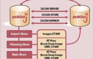
We have designed and built a web-based archive that enables users to view a large cross-section of images from a range of sources. Images and clinical data are contained in the site along with interpretations of the data from experienced users. The site contains mining tools that enable users to interrogate the archive. For example, ‘Show me images of those subjects in the archive that have a specific clinical presentation’. Alternatively the site allows users, through software bound into the site, to search for images with specific characteristics, for example uptake, shape and size.

Data mining

Presentation mining
I have a patient with a particular set of presentation characteristics. Show me images of a patient in the database with a similar set of characteristics.
The site has a series of windows that allow users to look at images with a specific presentation characteristic. One of these windows is shown in Figure 1.

Figure 1:
A window from the presentation mining section of fpcit.net.

 
Image mining
I have a set of patient images with a particular appearance. Show me images of patients in the database that look like this.
The site allows users to interrogate the imaging archive base on a number of imaging characteristics. For example show me images that have a caudate to background ratio of X calculated using a particular software approach. Figure 2 shows a particular software approach which comes as part of the Quantispect software and measures the ratio of the striatum to the occipital cortex.

Figure 2: The regions of interest used to quantify the striatum to background ratio.


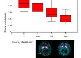
Semi quantitative techniques such as this do not produce ideal discrimination between Parkinsonian and non Parkinsonian patients. There is also some discrepancy between the visual interpretation and the semi-quantified value. Figure 3 shows a box plot of the visual interpretation and the quantified value.

Figure 3: Plot of semi quantified uptake and visual classification


The site also uses a pattern recognition technique to identify similar images. Image data can be uploaded to the site, processed on-line, and compared to the database. Figure 4 shows how the size and shape are extracted.

Figure 4: Extracting the size and shape from each striatum
NS/EW RATIO


Figure 5: Plot of shape measure and visual classification. The NS/EW ratio is the ratio if the length of the longest to the shortest axis.
SIZE AREA


The relationship between each shape measure and the visual classification is shown in Figure 5. Using a combination of shape and size and a nearest neighbor technique for classification the software had a kappa statistic of 0.91 when compared to an experienced observer.
Although this site is based around fpcit its structures and concept are generic for new imaging radiopharmaceutical techniques. fpcit.net provides a data archive and image processing tools that can aid any new users to ascend the learning curve. It is free, and has a powerful connectivity tools which enables it to be used with data acquired from any system.

By |Archive|Comments Off on fpcit.net: A web-based archive for use with 123I Ioflupane